Welcome to Sprint Diagnostics in Hyderabad, where your health and well-being are our topmost priority. In a world where healthcare is evolving rapidly, we stand firm in our commitment to offer you the best in diagnostic services. We combine cutting-edge technology, medical expertise, and a patient-centered approach to provide an unrivaled service in radiology diagnostics.
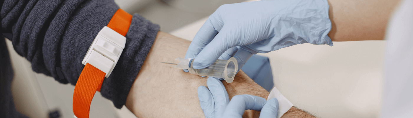
Home Sample Collection Process
1
Book your convenient slot
2
Sample Collection by Phlebotomist
3
Reporting of the sample at lab
4
Download Reports
Note: Home Sample Collection is only for Pathology lab tests.
We are proud to present the following features that make Sprint Diagnostics the preferred choice for your diagnostic needs:
State-of-the-Art Equipment: Our facility is equipped with the latest and most advanced diagnostic machines to deliver high-definition, accurate imaging results.
2
Precision-Based Radiology: Our precision-based approach, along with cutting-edge technologies, ensures that every diagnosis is precise, comprehensive, and reliable.
3
Advanced Technologies: We use the most innovative technologies in the field of radiology to offer you the highest standards of diagnostic services.
4
Experienced Doctors and Technicians: Our team comprises highly qualified and experienced doctors and technicians who are dedicated to providing the highest quality patient care.
5
Ambience: Our centre offers a serene and calming environment to ensure your comfort and well-being during the entire diagnostic procedure.
6
Prompt Reporting: We understand the importance of time in healthcare. Therefore, we ensure prompt reporting, so you receive your results as quickly as possible.
7
Affordability: We believe in making top-quality healthcare accessible. Our competitive pricing ensures everyone can afford our top-quality diagnostic services.
8
Quality First Motto: We stand by our "Quality First" motto. Our continuous efforts in improving our services ensure the best medical diagnostic experience.
9
Spectrum of Services: From MRI to CT, Ultrasound to Mammography, X-Ray to BMD, we offer a comprehensive range of diagnostic services under one roof.
10
Patient Education: We help patients understand their medical conditions and procedures to ensure they are fully aware and comfortable.
11
Easy Appointments: With our easy appointment booking system, you can plan your visit at your convenience.
12
Transparent Billing: Our transparent billing system ensures no hidden charges or surprises, aligning with our values of trust and honesty.
13
Constant Upgradation: We continually upgrade our equipment and technologies to keep up with the advancements in healthcare.
14
Community Engagement: We actively participate in community health programs, underlining our commitment to the broader well-being of society.
15
Infection Control: We adhere to strict infection control protocols to ensure a safe and sterile environment for our patients.
16
Comprehensive Health Packages: We offer comprehensive health packages designed to suit various health needs, encouraging preventive healthcare.
Experience the Sprint Diagnostics difference today! Your health deserves nothing but the best. So, why wait? Book your appointment now and let us help you on your journey towards better health.At Sprint Diagnostics, your health is not just our profession, it's our passion. We're not just providing diagnostic services; we're delivering peace of mind and paving the way for a healthier future. Your health deserves nothing but the best, and we promise to offer just that.