Tumor Scintigraphy
- Home
- Our Services
- Nuclear Medicine
- Gamma Camera
- Tumor Scintigraphy
Tumor scintigraphy is a diagnostic imaging procedure used to identify and monitor the growth of tumors within the body. This is achieved by using a small amount of radioactive substance, called a radiotracer, which can be detected by a special camera. This radiotracer is usually injected into a vein and is then absorbed by various body tissues. Tumor cells tend to absorb more of the radiotracer, and thus appear more brightly on the resulting images.
This diagnostic method is incredibly valuable to healthcare professionals because it enables the detection of cancerous tumors at an early stage, assesses the extent or stage of the cancer, monitors the efficacy of treatment, and even checks for recurrence after treatment. Furthermore, it provides crucial insights into how the tumor is affecting the body and how the body is responding to the presence of the tumor.
Home Sample Collection Process
Note: Home Sample Collection is only for Pathology lab tests.
Specific Instructions:
Before undergoing tumor scintigraphy, there are several key instructions that should be followed:
- Fasting : Depending on the type of scan, you may be required to fast for several hours prior to the procedure. Your doctor will provide specific instructions based on your health and the type of scan.
- Hydration : Unless directed otherwise, it's recommended to drink plenty of fluids to help flush the radiotracer from your body after the scan.
- Allergies : Inform your doctor about any known allergies, particularly any reactions to medications or iodine.
- Pregnancy and breastfeeding : If you are pregnant or breastfeeding, you should notify your doctor, as the radiation from the scan can pose risks to a developing fetus or a nursing child.
- Medications : Continue taking regular medications unless specifically told not to by your doctor. Also, let your doctor know about any over-the-counter drugs, supplements, or herbal remedies you are taking.
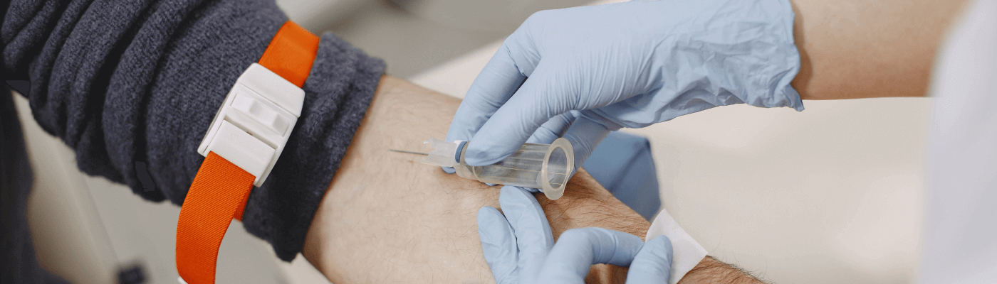
During the procedure, a radiotracer will be injected into a vein in your arm. You will then wait for the substance to travel through your body and be absorbed by the tissues. This waiting period can vary from a few minutes to a few hours. Once the radiotracer has had time to accumulate in the body tissues, you will be positioned on an examination table under a special camera that detects the radiation emitted by the radiotracer and produces images.
The procedure itself is generally not painful. The only discomfort you may experience is from the needle prick when the radiotracer is injected.
The total time can vary depending on the type of scan. The initial injection of the radiotracer takes only a few minutes, but the waiting time for the substance to circulate can vary from a few minutes to a few hours. The scan itself may take 30 minutes to an hour.
After the scan, you can usually return to your regular activities. Drinking plenty of fluids helps flush the radiotracer out of your system. The doctor will interpret the results and discuss them with you at a follow-up appointment.
Areas where the radiotracer is absorbed in greater amounts are called 'hot spots', and these often indicate the presence of cancerous tumors. Areas that absorb less tracer are called 'cold spots' and may indicate a lack of blood flow or tissue damage. Your doctor will interpret the images in relation to your symptoms and other diagnostic tests.
The risks are minimal. The amount of radiation you're exposed to is low and safe. However, there's a slight risk of an allergic reaction to the radiotracer, but this is rare. Always inform your doctor if you're pregnant, think you might be, or if you're breastfeeding.
The frequency of the test depends on the type of tumor, its stage, the treatment being given, and the doctor's assessment of your condition. Your doctor will provide guidance on how often the test should be repeated.
Various factors can affect the results of the test, including certain medications and substances, poor kidney function (as the radiotracer is often cleared by the kidneys), and inadequate preparation for the test, such as not fasting if required.
If your test results are abnormal, you should consult with an oncologist who specializes in the treatment of cancer.
No, tumor scintigraphy is just one of many tools that doctors use to diagnose and monitor cancer. Other diagnostic tools include other imaging tests like CT scans, MRIs, and PET scans, as well as biopsies and blood tests.
Tumor scintigraphy plays an important role in the diagnosis and management of cancer. It provides valuable information about the presence, location, and size of tumors, contributing to a comprehensive understanding of the patient's condition and guiding treatment decisions. As with any medical procedure, it is essential to be well-informed about the procedure, how to prepare for it, and what to expect during and after the scan. This knowledge can go a long way in relieving any anxiety related to the test and ensuring a smooth process.
- 4KM from Madhapur
- 3KM from Banjara Hills
- 1.9KM from Yusufguda
- 3KM from Madhura Nagar
- 5KM from Shaikpet